Breast ultrasound in Kyiv ✳️ prices for 2026
New Year holiday closure: 31.12.2025-04.01.2026
Kyiv, 72 Antonovycha St.
(Olimpiiska metro station)
Mon-Sat: 10:00 - 20:00
Sun: closed
Ultrasound scan

Breast ultrasound

Early detection of serious breast conditions will enable you to overcome the condition quickly and, in many cases, without the need for surgery.

Home / Services / Ultrasound scan / Breast ultrasound

01

Detection of pathological changes
from 4 mm

02

Without prior
training

03

Pain-free
and non-invasiveness

04

Absence
contraindications

05

Quick
result

06

The possibility of frequent
organisation

About the breast ultrasound procedure

Breast ultrasound is an accessible diagnostic method recommended for detecting abnormalities at any stage of development. The incidence of breast diseases is rising every year and requires timely detection and treatment to preserve women’s lives and health. Benign growths are diagnosed in one in three women, and cancer in one in eight.

What does a breast ultrasound examine?

Breast examinations are carried out on women of all ages. In just a few minutes, the doctor examines:

  • the nipple and areola;
  • glandular and connective tissue;
  • mammary ducts;
  • fascia;
  • pre-mammary and retro-mammary adipose tissue.

During a breast ultrasound, the nearby lymph nodes are also examined.

Play Video

Benefits of the procedure

Breast ultrasound is a completely non-invasive and safe method for diagnosing breast conditions. Unlike mammography, ultrasound does not expose the body to radiation, so it poses no risk to the patient’s health, has very few contraindications, and can be performed as often as necessary and dictated by the clinical situation.

  • Simple cysts – anechoic formations with a thin wall and no septa
  • Complex cysts – echogenic with thick internal walls
  • Benign lesions – round, encapsulated, anechoic or hyperechoic, without a vascular network
  • Suspected malignant tumours – hypoechoic, with irregular margins, without a capsule, and may contain calcifications
  • A fibroadenoma is a benign tumour with clear borders, round or oval in shape, hypoechoic, and without signs of encapsulation
Indications and contraindications for the service
Indications
Contraindications
  • Pain, which may be localised or affect the entire breast.
  • The appearance of discharge from the nipples — watery, bloody or greenish.
  • Galactorrhoea — the secretion of milk outside of breastfeeding.
  • Changes in the size and shape of the breast, nipple and areola.
  • The presence of lumps detected during a self-examination or a medical examination.
  • Enlargement of the nearby lymph nodes.
  • The appearance of hardened areas around the collarbones and in the armpits.
  • Breast tenderness not associated with the first half of the menstrual cycle.
Contraindications
  • Open wounds or injuries in the breast area that prevent the sensor from making contact with the skin.
  • Purulent or other infectious processes in the area under examination.
  • Damage to the skin around the breasts (burns, severe rashes).
  • The patient’s psychological or physical inability to remain still during the procedure.

Important! An ultrasound scan of the breasts is recommended as a matter of urgency in cases of injury involving bruising, scratches or contusions to the chest. The scan is also recommended for young women before they start taking oral contraceptives.

Request a consultation

Leave a request for a consultation

How should I prepare for a breast ultrasound scan?

No special preparation is usually required for a breast ultrasound scan. However, there are some important points to bear in mind when planning an ultrasound examination. The ideal time for an ultrasound scan is between the 5th and 12th day of the menstrual cycle for women of reproductive age. During this period, breast tissue is less dense, which facilitates visualisation and allows for better interpretation of the results.

For women who are postmenopausal (or taking hormonal contraceptives), the day of the menstrual cycle is irrelevant for performing an ultrasound scan.

What does an ultrasound scan involve?

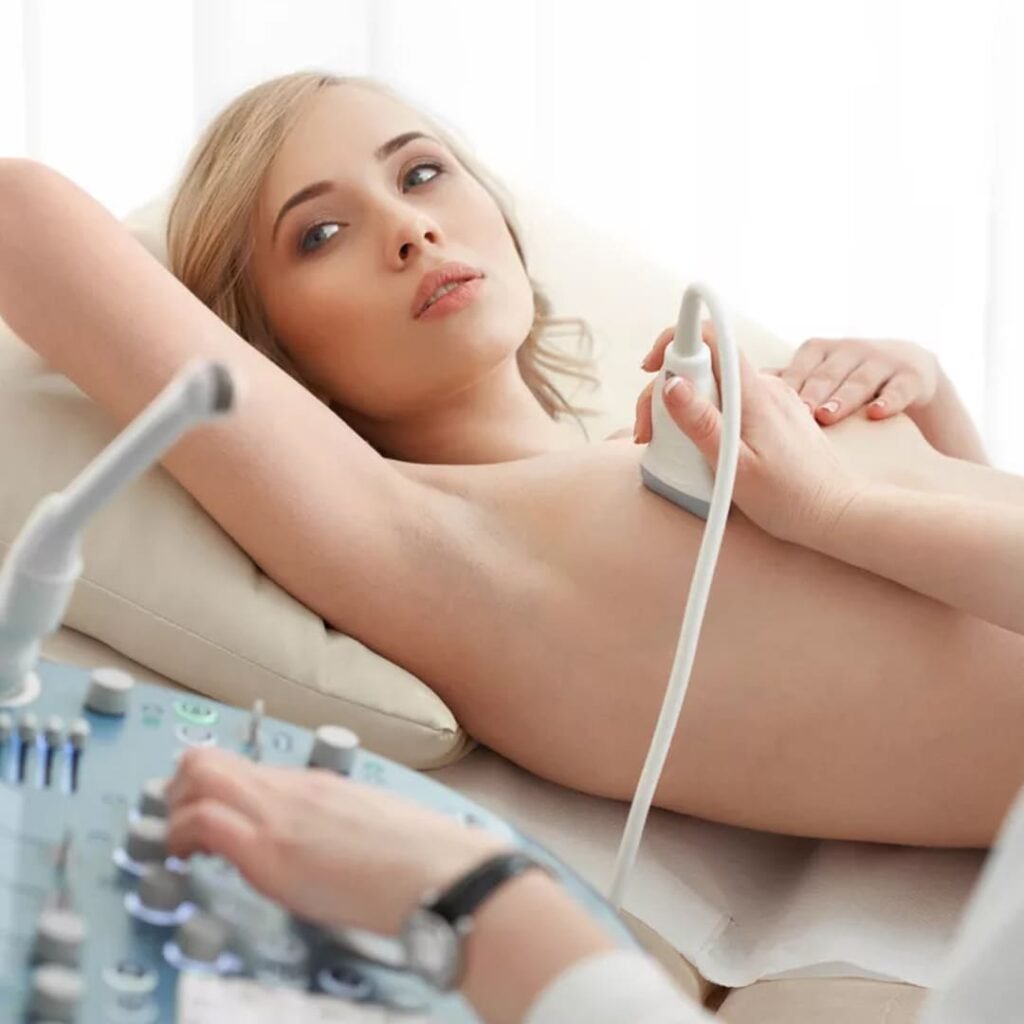
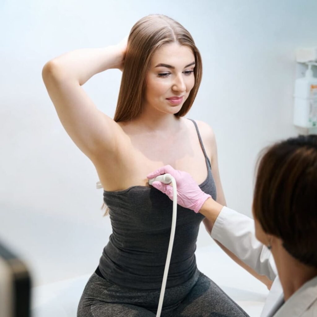
The patient lies on her back on a comfortable examination couch. Depending on the angle required to view the area being examined, the doctor may ask her to turn onto her side or sit up. A special gel is applied to the breast area to improve contact between the transducer and the skin, eliminating air pockets that could distort the ultrasound waves.

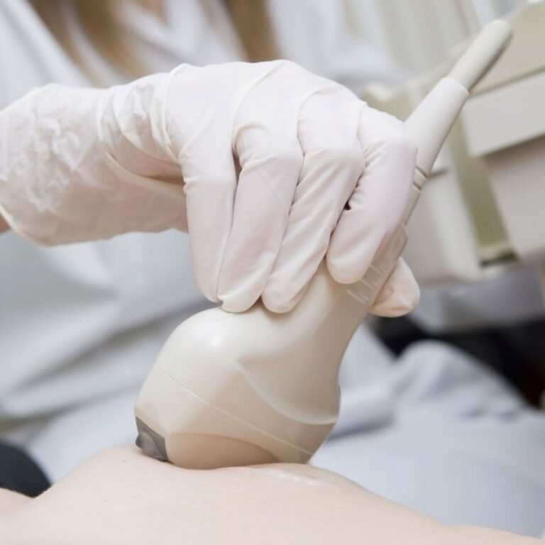
The doctor gently moves the probe across the breasts, obtaining images of the internal structures. The patient may feel slightly uncomfortable due to the pressure of the probe, but the procedure is not painful.

During the ultrasound scan, the doctor assesses the homogeneity of the tissue, the presence and nature of lumps, cysts, nodules and other abnormalities. The size and characteristics of any lesions found, as well as the condition of the lymph nodes and blood flow in the area of the lesions, are also assessed during the examination.

  • The procedure takes 15–20 minutes
  • The results are available immediately after the examination

How do I get the results?

You can receive the results of your breast ultrasound within 10–15 minutes of the examination being completed. These include:

  • images showing various structures and any possible changes

  • a detailed written report from the diagnostic radiologist, which includes:

  • a description of the homogeneity of breast tissue, and the presence and nature of any lumps, cysts, nodules and other structures;

  • the dimensions and characteristics of any lesions, if detected;

  • an assessment of the condition of the lymph nodes in the axillary region;

  • an assessment of blood flow in the area of the detected lesions (if a Doppler scan was performed rather than a standard scan);

  • an interpretation of the ultrasound findings, indicating possible pathologies and recommendations for further investigation or treatment.

If any abnormalities are detected, your doctor may recommend a mammogram, an MRI scan, a biopsy or a consultation with a breast specialist for a more thorough assessment and to determine the next steps in your treatment or monitoring.

Our team of doctors

BG-Lab doctors who provide this service

Obstetrician-Gynecologist-Endocrinologist, Ultrasound Specialist, Specialist in Aesthetic Gynecology and Laser Techniques, Nutritionist, Pellet Therapy Specialist

Gynecologist-Endocrinologist, Ultrasound Specialist, Aesthetic Gynecology and Laser Expert, Pellet Therapy Specialist, Sexologist

Breast ultrasound scan price
  • Consultation with a gynaecologist
1000 UAH
  • Online consultation with a gynaecologist
1000 UAH
  • Online consultation with an endocrinologist
1000 UAH
  • ‘Women’s Confidence’ Check-up (three ultrasound scans, consultation with a gynaecologist-endocrinologist)
4050 UAH 1500 UAH
*We are offering a 63% discount on a gynaecological check-up with a doctor who has over 20 years’ experience
  • ‘Women’s Confidence+’ Check-up (eight ultrasound scans, consultation with a gynaecologist-endocrinologist)
2500 UAH
  • Consultation with a gynaecologist and aesthetic specialist, PhD in Medical Sciences
1200 UAH
  • Breast ultrasound
500 UAH

Other services

Author of the article: Yulia Vitaliivna Lezhnova – ultrasound specialist, gynaecologist, endocrinologist

Reviewer of the article: Tetiana Vladyslavivna Tsurkan – Chief Physician, international expert, dermatologist, speaker at international dermatology congresses on applied aesthetics and cosmetology